Grey or white – does it matter?
advertisement
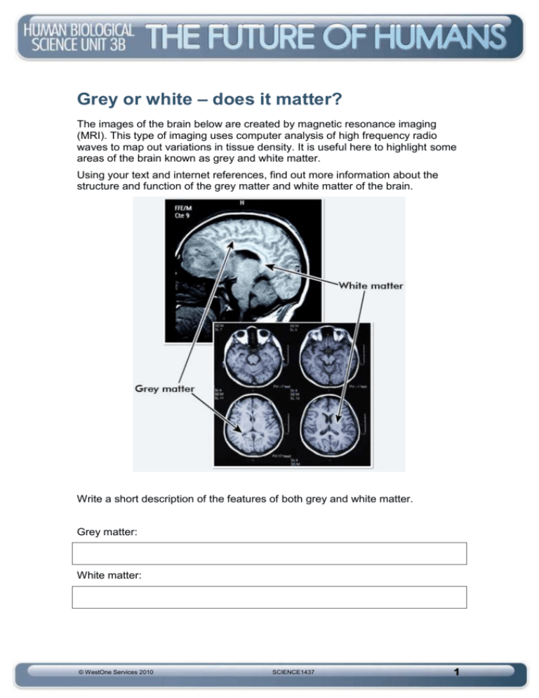
Grey or white – does it matter? The images of the brain below are created by magnetic resonance imaging (MRI). This type of imaging uses computer analysis of high frequency radio waves to map out variations in tissue density. It is useful here to highlight some areas of the brain known as grey and white matter. Using your text and internet references, find out more information about the structure and function of the grey matter and white matter of the brain. Write a short description of the features of both grey and white matter. Grey matter: White matter: © WestOne Services 2010 SCIENCE1437 1 The cerebral hemispheres The outer few millimetres of the two cerebral hemispheres is known as the cerebral cortex. Humans have the largest cerebral cortex of all primates. It is folded in patterns called convolutions or gyri that give it a greatly increased surface area. The convolutions are separated by shallow depressions called sulci and deep depressions called fissures. Describe the difference between these three features. The cerebrum The left and right cerebral hemispheres are divided by the longitudinal fissure and can then be further subdivided into four lobes – the frontal, parietal, occipital and temporal. (Note: There is a fifth lobe, the insula, but this is deep within the brain) Using your text and other references shade the four different lobes of the brain, labelling each one and include a brief description of what each one controls. © WestOne Services 2010 SCIENCE1437 2 The four lobes of the cerebral hemispheres are: the ______________________ which controls ____________________________________ the ______________________which controls _____________________________________ the ______________________ which controls ____________________________________ the______________________ which controls ____________________________________ © WestOne Services 2010 SCIENCE1437 3 Functions of the cerebrum 1. Use your text and other references to label the following important structures and regions on the diagram below: primary sensory area primary motor area visual association area primary visual association area primary auditory area auditory association area primary speech area. 2. Use colours to identify the sensory, motor and association areas of the brain. Sensory areas receive impulses from the senses. Motor areas send impulses to the muscles for movement. Association areas interpret and process information relating to intellect and emotions. 3. Now write the functions of each of the following: primary sensory area © WestOne Services 2010 SCIENCE1437 4 primary motor area visual association area primary visual association area primary auditory area auditory association area primary speech area © WestOne Services 2010 SCIENCE1437 5
