Medical Imaging Quiz − Case 21
advertisement

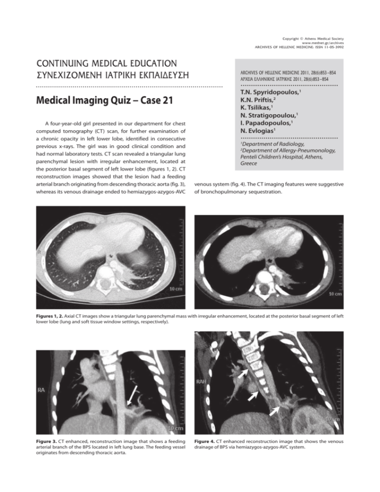
Copyright © Athens Medical Society www.mednet.gr/archives 853 ARCHIVES OF HELLENIC MEDICINE: ISSN 11-05-3992 MEDICAL IMAGING QUIZ – CASE 21 CONTINUING MEDICAL EDUCATION ΣΥΝΕΧΙΖΟΜΕΝΗ ΙΑΤΡΙΚΗ ΕΚΠΑΙ∆ΕΥΣΗ ARCHIVES OF HELLENIC MEDICINE 2011, 28(6):853-854 ÁÑ×ÅÉÁ ÅËËÇÍÉÊÇÓ ÉÁÔÑÉÊÇÓ 2011, 28(6):853-854 ............................................... Medical Imaging Quiz − Case 21 A four-year-old girl presented in our department for chest computed tomography (CT) scan, for further examination of a chronic opacity in left lower lobe, identified in consecutive previous x-rays. The girl was in good clinical condition and had normal laboratory tests. CT scan revealed a triangular lung parenchymal lesion with irregular enhancement, located at the posterior basal segment of left lower lobe (figures 1, 2). CT reconstruction images showed that the lesion had a feeding arterial branch originating from descending thoracic aorta (fig. 3), whereas its venous drainage ended to hemiazygos-azygos-AVC T.N. Spyridopoulos,1 K.N. Priftis,2 K. Tsilikas,1 N. Stratigopoulou,1 I. Papadopoulos,1 N. Evlogias1 ............................................... 1 Department of Radiology, Department of Allergy-Pneumonology, Penteli Children’s Hospital, Athens, Greece 2 venous system (fig. 4). The CT imaging features were suggestive of bronchopulmonary sequestration. Figures 1, 2. Axial CT images show a triangular lung parenchymal mass with irregular enhancement, located at the posterior basal segment of left lower lobe (lung and soft tissue window settings, respectively). Figure 3. CT enhanced, reconstruction image that shows a feeding arterial branch of the BPS located in left lung base. The feeding vessel originates from descending thoracic aorta. Figure 4. CT enhanced reconstruction image that shows the venous drainage of BPS via hemiazygos-azygos-AVC system. 854 Bronchopulmonary sequestration (BPS) is a congenital area of dysplastic and nonfunctioning lung which is not connected with the bronchial tree or the pulmonary arteries. Other congenital anomalies may also coexist (heart, musculoskeletal system etc.). The appearance of BPS on prenatal ultrasound examination is similar to that of microcystic congenital cystic adenomatoid malformation. However, BPS has a systemic arterial blood supply; a color flow Doppler ultrasound showing a systemic supply to the mass could confirm the diagnosis. The feeding branch of the abnormal lung area typically arises from descending aorta. BPS is most commonly located in left lower lobe, followed by right lower lobe.1,2 BPS may be either intralobar (75%) which is enclosed by visceral pleura of affected pulmonary lobe but separated from the bronchial tree or extralobar (25%), which is actually an accessory lobe with its own pleural sheath.1,2 Intralobar BSP is acquired and commonly seen among adults (50% >20 years) and usually presents with recurrent acute lower lobe pneumonias (asymptomatic in about 15% of cases). It is a homogenous or inhomogenous soft tissue mass with irregular borders. Mucoid impaction of dilated bronchus surrounded by hyperinflated lung is characteristic of such lesion, which may also include thin wall cysts, with air-fluid levels in case of infection.3,4 Extralobar BSP is congenital and most frequently seen among infants and small children. It is usually a single, well defined soft tissue triangular mass, most commonly located adjacent to the posterior medial hemidiaphragm.3,4 Radiographic images play a fundamental role in establishing the diagnosis, and in providing the medical team with a vascular map for surgical planning.5 References 1. CORBETT HJ, HUMPHREY GM. Pulmonary sequestration. Pediatr Respir Rev 2004, 5:59−68 2. BOLCA N, TOPAL U, BAYRAM S. Bronchopulmonary sequestration: Radiologic findings. Eur J Radiol 2004, 52:185−191 3. AHMED M, JACOBI V, VOGL TJ. Multislice CT and CT angiography for non-invasive evaluation of bronchopulmonary sequestration. Eur Radiol 2004, 14:2141−2143 4. LEE EY, SIEGEL MJ, SIERRA LM, FOGLIA RP. Evaluation of angioarchitecture of pulmonary sequestration in pediatric patients using 3D MDCT angiography. AJR Am J Roentgenol 2004, 183:183−188 5. ABBEY P, DAS CJ, PANGTEY GS, SEITH A, DUTTA R, KUMAR A. Imaging in bronchopulmonary sequestration. J Med Imaging Radiat Oncol 2009, 53:22−31 Corresponding author: T.N. Spyridopoulos, Department of Radiology, Penteli Children’s Hospital, Athens, Greece, tel.: +30 2132 052 569 e-mail: thspyrid@med.uoa.gr Diagnosis: Bronchopulmonary sequestration Comment T.N. SPYRIDOPOULOS et al ..............................................................................................................................


